

FRCR anatomy is the Part 1 module of 100 images in 90 minutes, each with one arrowed structure to name, so you have under a minute per image and precise naming earns full marks. Prepare by seeing a high volume of images across modalities and regions, and by practising exact, side-specific answers against the clock.
This guide covers the format and marking, what to cover, naming and laterality, an eight-week plan, a practice method, common traps and tactics for the day.
Table of Contents
How the FRCR anatomy module is set out
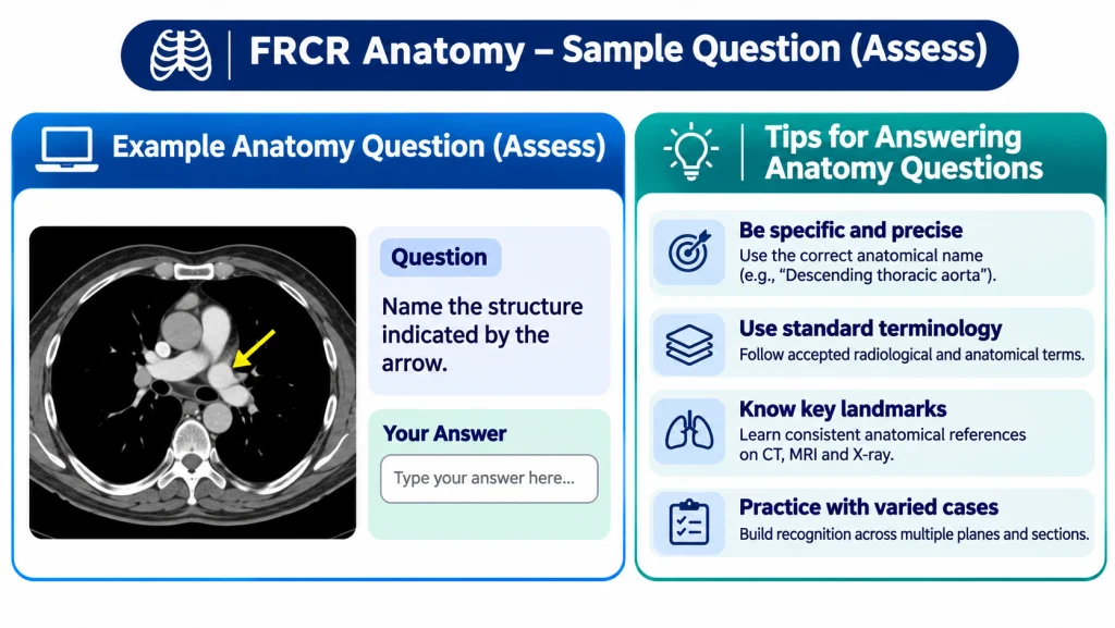
The anatomy module shows 100 images in 90 minutes on Assess, the RCR’s image-viewing platform. On each image, one structure is marked by one or more arrows, and there is a single question about it. You type a short answer, then move on.
Marking rewards accuracy. A completely accurate answer scores two marks, a less accurate but still correct answer scores one, and an incorrect answer scores zero. There is no negative marking, so an attempt never costs you marks. Answers that do not exactly match the platform’s accepted answers are reviewed by examiners, which means a sensible answer is not lost to a trivial mismatch, but you should not rely on that review to rescue vague terms.
| Feature | FRCR anatomy module |
| Images | 100, each with one arrowed structure |
| Time | 90 minutes, about 54 seconds per image |
| Platform | Assess |
| Marking | +2 completely accurate, +1 less accurate but correct, 0 incorrect |
| Negative marking | None |
| Pass mark | Modified Angoff, finalised after the exam by the Hofstee method |
Anatomy is one of the two First FRCR modules with physics. You can sit them together or separately, in either order, as explained in our FRCR Part 1 exam guide.
Image types and regions to cover
Cover the whole body across the imaging modalities you report in daily practice. The RCR does not publish a breakdown of anatomy images by region or modality, so do not plan around a guessed weighting. Treat every region as examinable and use the RCR’s anatomy advice document, listed under candidate resources on the CR1 exam page, as your official reference.
In practical terms, that means working through head and neck, neuroanatomy, chest and cardiovascular, abdomen and pelvis, the spine and the musculoskeletal system. Within each region, look at the structure on plain radiographs, cross-sectional CT and MRI, and ultrasound where relevant, because the same structure looks quite different from one modality to the next.
Do not forget the platform itself. The RCR provides an interactive demonstration of Assess. Spend time with it so that moving through images and entering answers feel familiar before the exam.
Naming precision, spelling and laterality
Precision is where FRCR anatomy marks are won or lost. Since full marks go to a completely accurate answer, name the exact structure the arrow points to, not its parent or region. If the arrow sits on a specific branch, a specific nerve root or a specific part of a bone, say so.
State the side whenever a structure is paired. Get into the habit of checking orientation markers and the image’s left and right before you answer, especially on axial images where it is easy to reverse sides under pressure. Naming the right structure on the wrong side risks losing the marks for that image.
Spell terms properly and use standard anatomical names. Examiners review answers that do not exactly match the accepted list, but clear, correctly spelled terminology is the safest route to two marks. Avoid abbreviations unless they are universally standard, and avoid eponyms unless you are sure they are accepted.
An 8-week FRCR anatomy plan
This eight-week plan assumes around eight to ten hours a week alongside a full rota, built around daily image sets. If you are sitting physics at the same sitting, run the two in parallel as described in our FRCR Part 1 preparation guide.
| Week | Regions | Daily practice |
| 1 | Head and neck, skull base | 25 images, untimed, check every answer |
| 2 | Neuroanatomy | 30 images, start a naming log |
| 3 | Chest, mediastinum and heart | 30 images, begin timing sets |
| 4 | Vascular anatomy across the body | 40 images at about a minute each |
| 5 | Abdomen and pelvis | 40 timed images, revisit naming log |
| 6 | Spine and upper limb | 50 timed images |
| 7 | Lower limb, mixed regions | One full 100-image mock in 90 minutes |
| 8 | Weak regions, second full mock | Light review in the last two days |
Mix modalities within every week rather than doing all radiographs one week and all MRI the next. The anatomy module will not group images for you.
A practice method that builds speed
FRCR anatomy practice should start slow and accurate, then get faster. In the first weeks, take as long as you need on each image, check the answer and note the exact term in a naming log. Accuracy built early becomes speed later.
From week three onwards, time every set. Aim for about a minute per image at first, then tighten towards the real pace of 54 seconds. When you get an image wrong, write down why: wrong structure, wrong side, imprecise term or misspelling. Those four categories are common ways marks slip away, and they tell you exactly what to fix.
Use your daily reporting as extra practice. When you scroll through a CT or MRI at work, pause on a few structures and name them precisely with their side. If you want structured support, StudyFRCR has an FRCR Part 1 course; see the course page for what it includes.
Common traps in the anatomy module
The commonest traps in the anatomy module are about precision and pace rather than knowledge. Naming the region instead of the structure, forgetting laterality and reversing sides on axial images all cost marks you could have kept.
Other traps are worth knowing. Spending two or three minutes on one hard image steals time from easier ones later. Leaving an image blank throws away a possible mark when there is no negative marking.
Revising only from labelled atlases means you see structures in ideal views, not on the real clinical images the exam uses. And skipping the Assess demonstration leaves you learning the viewer on the day.
Tactics on the day
On the day, keep a steady rhythm of under a minute per image and never leave an answer blank. If an image stumps you, give your best specific answer, flag it if the platform allows, and move on.
Before you type, run a quick check: what is the arrow on, which side is it, and what is the most specific accurate name? That habit, practised for eight weeks, is what turns knowledge into marks. Anatomy falls the day after physics in each sitting, so plan rest between the two; dates are on our FRCR exam dates page.
If you are travelling, build that into your plan too. Hyderabad is the only Indian venue for CR1, and no Gulf venue is listed, so doctors in the UAE, Qatar or Saudi Arabia need to travel to Hyderabad or another listed venue. Arrive a day early, carry your passport and keep the evening before the FRCR anatomy paper quiet.
Frequently asked questions
How many images are in FRCR anatomy?
There are 100 images in 90 minutes, each with one structure marked by an arrow or arrows and a single question about it.
How is the anatomy module marked?
A completely accurate answer scores two marks, a less accurate but still correct answer scores one, and an incorrect answer scores zero. There is no negative marking.
Does spelling matter in FRCR anatomy?
Answers that do not exactly match the accepted list are reviewed by examiners, but correct spelling and standard terms are the safest way to score full marks.
Should I always state the side?
Yes, for any paired structure. Check orientation before answering, because naming the right structure on the wrong side risks losing the marks.
Does the RCR publish which regions are tested most?
No breakdown by region or modality is published for anatomy, so prepare the whole body across modalities and use the RCR’s anatomy advice document.
Can I practise on the exam platform?
Yes. The RCR offers an interactive demonstration of Assess on its website, and it is worth using well before the exam.
Ready to sharpen your image naming? Explore the FRCR Part 1 course or Talk to our FRCR team.
Related guides: FRCR Part 1 exam format · FRCR physics guide · FRCR Part 1 preparation · FRCR exam dates
Sources: RCR: FRCR Part 1 Radiology (CR1) · RCR: CR1 guidance notes for candidates · RCR: Exam regulations and policies